Phone
+33 (0)1 76 54 08 64
Address
66 avenue des Champs-Élysées
75008 Paris – France
Authors: Magalie Bénard, Damien Schapman, Christophe Chamot, Fatéméh Dubois, Guénaëlle Levallet, Hitoshi Komuro and Ludovic Galas
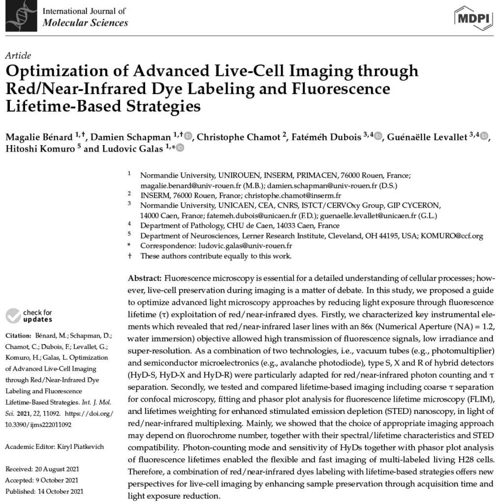
Abstract: Fluorescence microscopy is essential for a detailed understanding of cellular processes; how- ever, live-cell preservation during imaging is a matter of debate. In this study, we proposed a guide to optimize advanced light microscopy approaches by reducing light exposure through fluorescence lifetime (𝓣)exploitation of red/near-infrared dyes. Firstly, we characterized key instrumental elements which revealed that red/near-infrared laser lines with an 86x (Numerical Aperture (NA) = 1.2, water immersion) objective allowed high transmission of fluorescence signals, low irradiance and super-resolution. As a combination of two technologies, i.e., vacuum tubes (e.g., photomultiplier) and semiconductor microelectronics (e.g., avalanche photodiode), type S, X and R of hybrid detectors (HyD-S, HyD-X and HyD-R) were particularly adapted for red/near-infrared photon counting and 𝓣 separation. Secondly, we tested and compared lifetime-based imaging including coarse 𝓣 separation for confocal microscopy, fitting and phasor plot analysis for fluorescence lifetime microscopy (FLIM), and lifetimes weighting for enhanced stimulated emission depletion (STED) nanoscopy, in light of red/near-infrared multiplexing. Mainly, we showed that the choice of appropriate imaging approach may depend on fluorochrome number, together with their spectral/lifetime characteristics and STED compatibility. Photon-counting mode and sensitivity of HyDs together with phasor plot analysis of fluorescence lifetimes enabled the flexible and fast imaging of multi-labeled living H28 cells. Therefore, a combination of red/near-infrared dyes labeling with lifetime-based strategies offers new perspectives for live-cell imaging by enhancing sample preservation through acquisition time and light exposure reduction.
LBL-Dye Mito 715 formerly known as LBL-Dye M715 in this publication
LBL-Dye CellTag 717 formerly known as LBL-Dye M717 in this publication

The efficacy of fluorescence-guided surgery in facilitating the real-time delineation of tumours depends on the optical contrast of tumour tissue over healthy tissue. Here we show that CJ215—a commercially available, renally cleared carbocyanine dye sensitive to apoptosis, and with an absorption and emission spectra suitable for near-infrared fluorescence imaging (wavelengths of 650–900 nm) and shortwave infrared (SWIR) fluorescence imaging (900–1,700 nm)—can facilitate fluorescence-guided tumour screening, tumour resection and the assessment of wound healing...
The nuclear receptor, Nurr1, is critical for both the development and maintenance of midbrain dopamine neurons, representing a promising molecular target for Parkinson’s disease (PD)...
Cell-to-cell communication via tunneling nanotubes (TNTs) is a challenging topic with a growing interest. In this work, we proposed several innovative tools that use red/near-infrared dye labeling and employ lifetime-based imaging strategies to investigate the dynamics of TNTs in a living mesothelial H28 cell line that exhibits spontaneously TNT1 and TNT2 subtypes...
Proimaging