Phone
+33 (0)1 76 54 08 64
Address
66 avenue des Champs-Élysées
75008 Paris – France


Studying mitochondrial dynamics within tunneling nanotubes (TNTs) through live cell imaging presents several challenges and requires sophisticated techniques to capture the intricacies of these dynamic structures. Combining advanced imaging techniques such as fast FLIM (Fluorescence Lifetime Imaging Microscopy), confocal microscopy, and STED (Stimulated Emission Depletion) nanoscopy can offer valuable insights into the behavior of mitochondria within TNTs at high spatial and temporal resolutions.
By integrating these advanced imaging techniques, researchers can investigate various aspects of mitochondrial dynamics within TNTs, including mitochondrial transport, fusion-fission events, and interactions with other organelles or molecules. Additionally, live cell imaging enables the observation of real-time changes in mitochondrial behavior within TNTs, shedding light on the functional significance of these intercellular communication channels in cellular physiology and pathology.
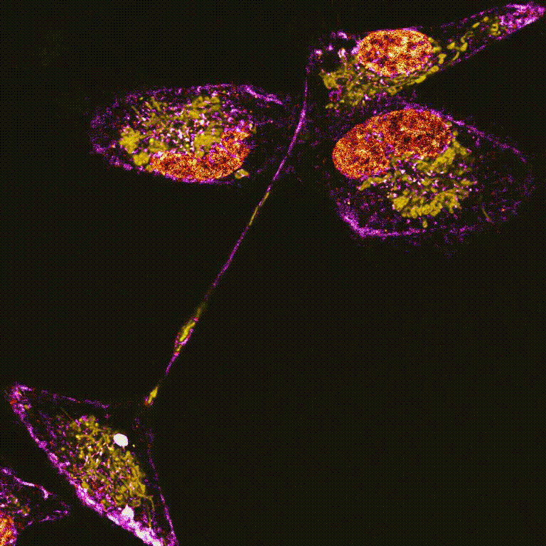
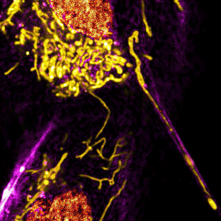
Images from the publication "Combining sophisticated fast FLIM, confocal microscopy, and STED nanoscopy for live-cell imaging of tunneling nanotubes"

Dynamics of mitochondria labeled with LBL-Dye Mito 715 in H28 cells during a 3-min time-lapse of lifetime dye unmixing overlayed images (1 image/3 sec) obtained through FLIM-integrated confocal microscopy.

Overlays of lifetime dye unmixing images showing the dynamics of mitochondria labeled with LBL-Dye Mito 715 in H28 cells during a 3-min time-lapse. White dotted line delimitated cell body and associated-TNT1
Dynamics of mitochondria labeled with LBL-Dye Mito 715 within a TNT1 connecting H28 cell bodies during a 10-min time-lapse of lifetime dye unmixing overlayed images (1 image/5 sec) obtained through FLIM-integrated confocal microscopy.

Time-lapse acquisition (10 min, 1 image/5 sec) in regions of interest closed to Cell #1 (A2 - A5) and Cell #2 bodies (A6 - A9) after mitochondria labeling with LBL-Dye Mito 715 Short light blue and green arrows indicated mitochondria migration and membrane bulging/mitochondria reorientation respectively.
Dynamics of mitochondria labeled with LBL-Dye Mito 715 within a TNT1 during a 20-min time-lapse of lifetime dye unmixing overlayed images (1 image/30 sec) obtained through FLIM-integrated confocal microscopy.

LBL-Dye Mito 715 allowing to follow mitochondrial dynamics (20 min time-lapse – Video 3, 1 image/30 sec, orange arrows) including fission and bilateral migration (red arrows) within TNT1 and cell body. Reference red dotted lines facilitated the observation of positions during migration.

Dynamics of mitochondria labeled with LBL-Dye Mito 715 within TNT1 during a 20-min time-lapse of lifetime denoising 3D-reconstructed Maximum Intensity Projection (MIP) images (z-stack; 1 image/30 sec) obtained through FLIM-integrated STED nanoscopy.

Dynamics of LBL-Dye Mito 715 labeled-mitochondria within TNT1 connecting live H28 cells through STED imaging and lifetime denoising. LBL-Dye M715 signal (ex 690 nm, 3% WLL laser power – HyD-X 705-740 nm) was depleted with 2% of 775-nm pulsed laser and processed with lifetime denoising. (A) Time-lapse (20 min, 1 image/30 sec) showing the trajectory of 3 mitochondria (A2, A3 and A4) within TNT1 emerging from the Cell #1 body (arrows, A1-A4). (B) Following (A), time-lapse (20 min, 1 image/30 sec) showing the trajectory of 1 mitochondrion within TNT1 reaching the Cell #2 body. Graphs represented the distance (µm) from the origin to the end time points (min) of tracked objects. Trajectories (Fire LUT) were obtained by using Fiji software e.g. plugin Manual Tracking. White dotted line delimitated cell body and associated-TNT1.

The efficacy of fluorescence-guided surgery in facilitating the real-time delineation of tumours depends on the optical contrast of tumour tissue over healthy tissue. Here we show that CJ215—a commercially available, renally cleared carbocyanine dye sensitive to apoptosis, and with an absorption and emission spectra suitable for near-infrared fluorescence imaging (wavelengths of 650–900 nm) and shortwave infrared (SWIR) fluorescence imaging (900–1,700 nm)—can facilitate fluorescence-guided tumour screening, tumour resection and the assessment of wound healing...
The efficacy of fluorescence-guided surgery in facilitating the real-time delineation of tumours depends on the optical contrast of tumour tissue over healthy tissue. Here we show that CJ215—a commercially available, renally cleared carbocyanine dye sensitive to apoptosis, and with an absorption and emission spectra suitable for near-infrared fluorescence imaging (wavelengths of 650–900 nm) and shortwave infrared (SWIR) fluorescence imaging (900–1,700 nm)—can facilitate fluorescence-guided tumour screening, tumour resection and the assessment of wound healing...
The efficacy of fluorescence-guided surgery in facilitating the real-time delineation of tumours depends on the optical contrast of tumour tissue over healthy tissue. Here we show that CJ215—a commercially available, renally cleared carbocyanine dye sensitive to apoptosis, and with an absorption and emission spectra suitable for near-infrared fluorescence imaging (wavelengths of 650–900 nm) and shortwave infrared (SWIR) fluorescence imaging (900–1,700 nm)—can facilitate fluorescence-guided tumour screening, tumour resection and the assessment of wound healing...
Proimaging