Phone
+33 (0)1 76 54 08 64
Address
66 avenue des Champs-Élysées
75008 Paris – France


Cellular tracers in fluorescence imaging are essential tools for studying various cellular processes, including cell migration, protein trafficking, organelle dynamics, and more...
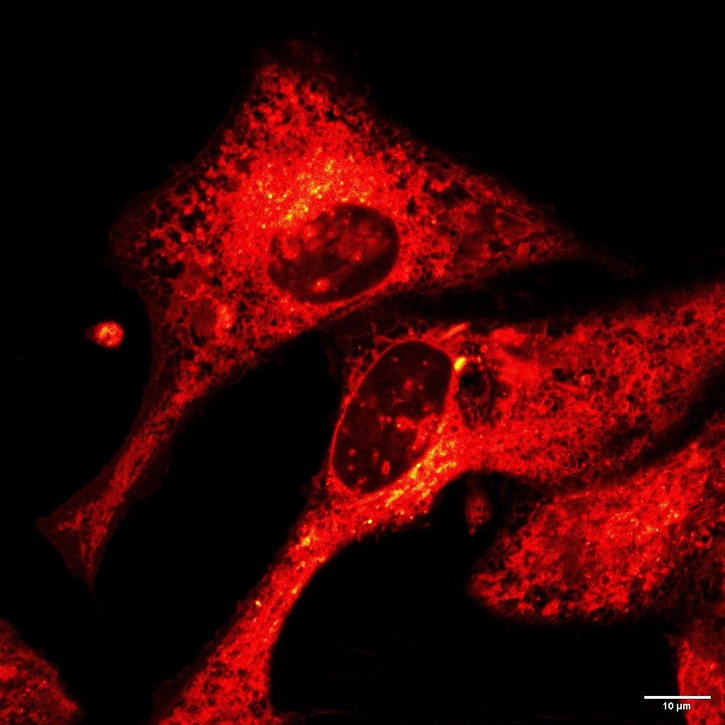
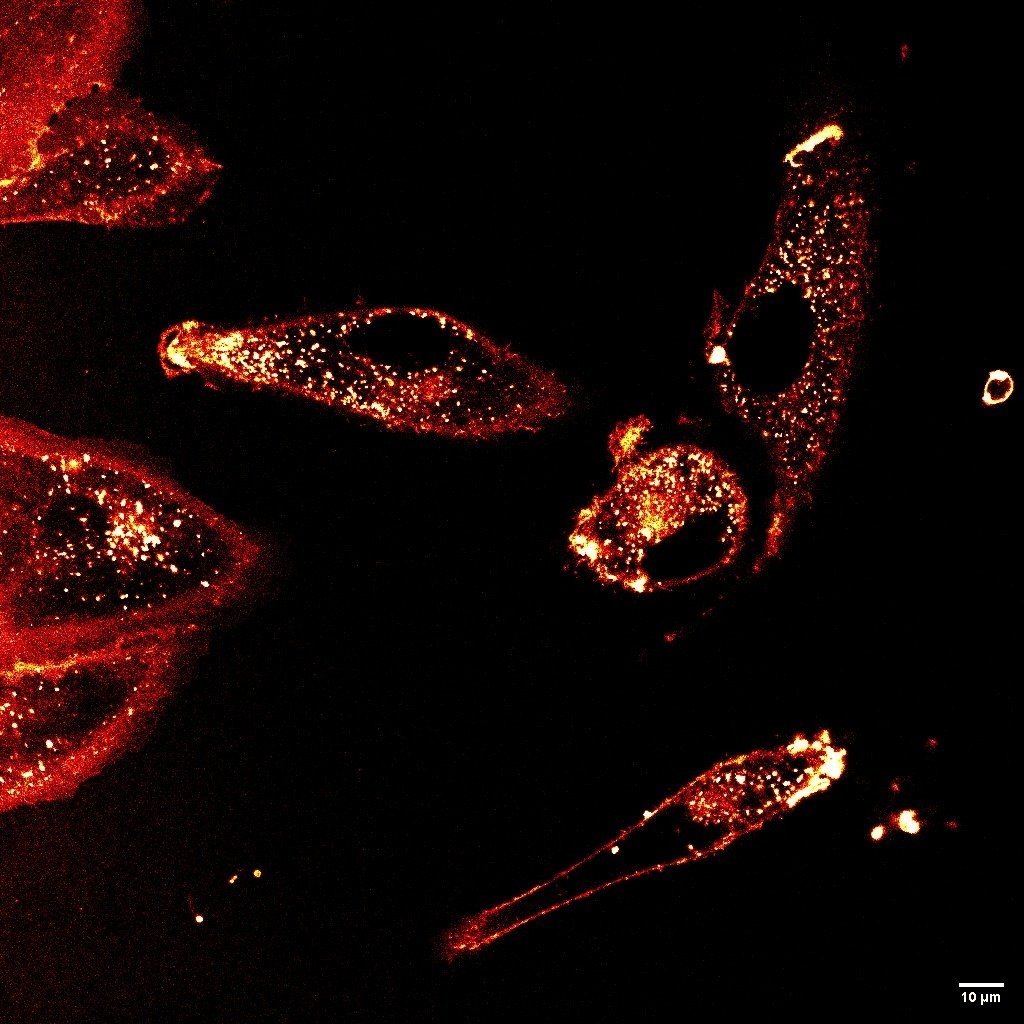
LBL-Dye CellTag 580 and LBL-Dye CellTag 717 (compatible with super-resolution STED imaging) are two cell-permeant, photostable, and no cytotoxic cellular tracers particularly well-adapted for long-time imaging on living cells.
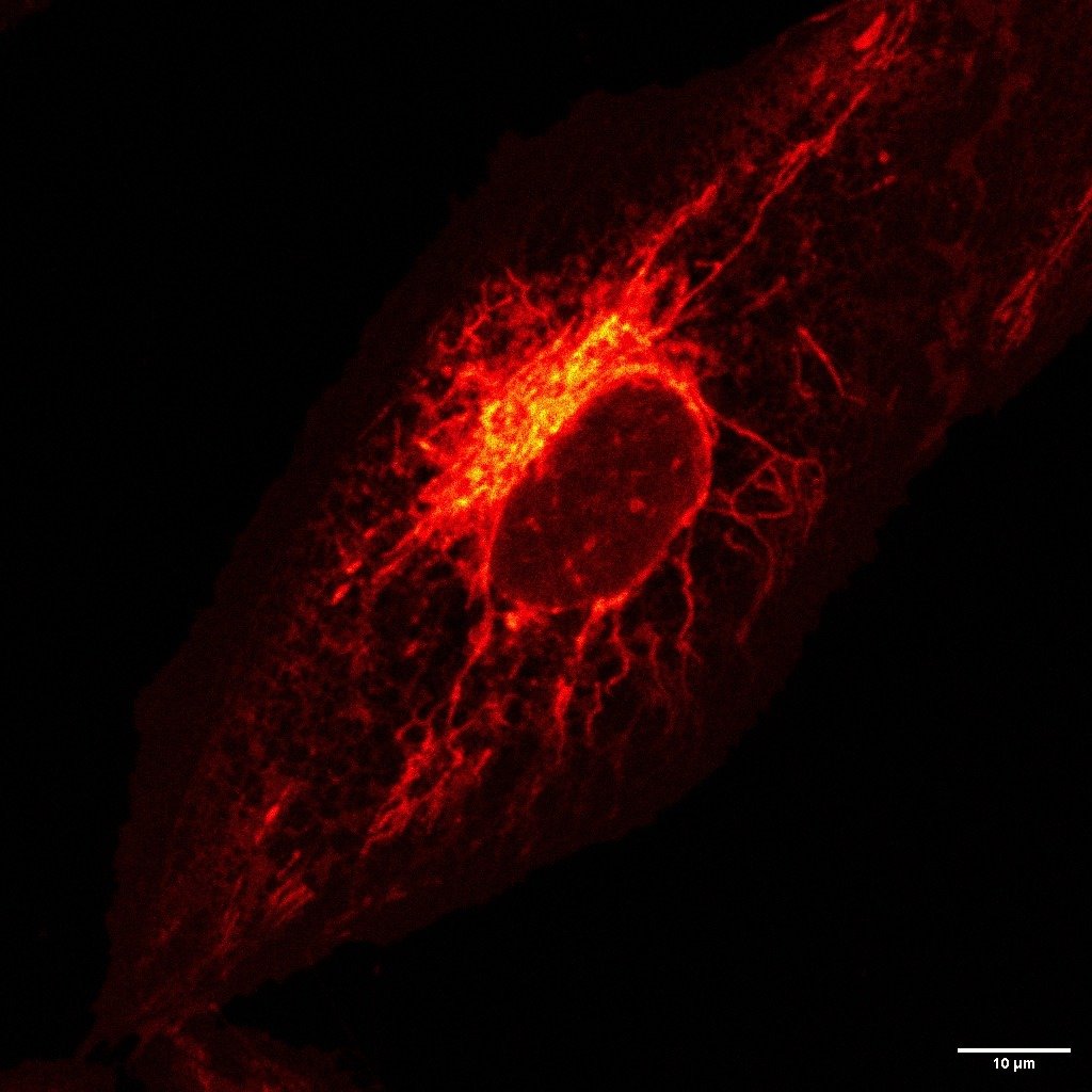
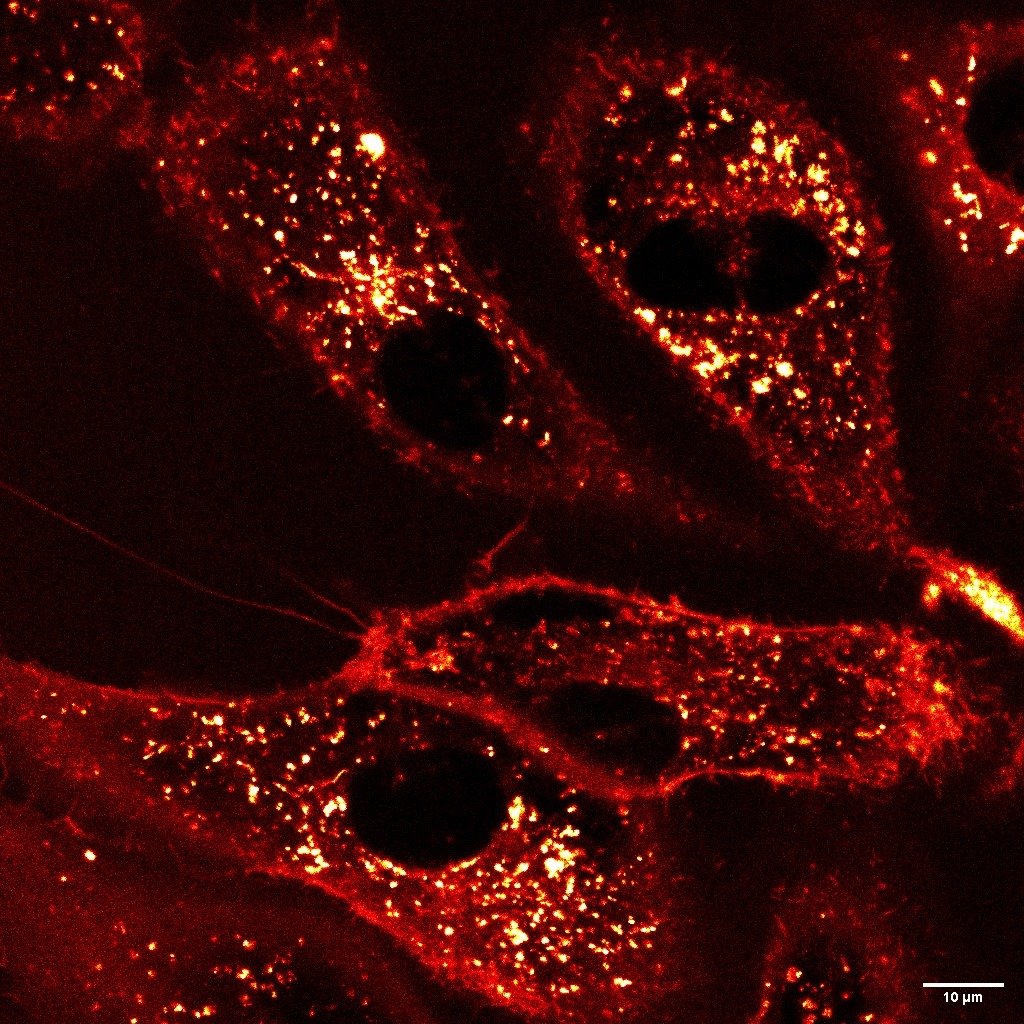
Imaging of H28-living cells stained with far-red LBL-Dye CellTag 580 (2µg/µl) in the culture medium. Images acquired by using STELLARIS8 confocal microscope (Leica Microsystems), X86 objective water immersion, WLL laser excitation 562 nm; Em 570-650 nm on HyD-S detector, Okolab chamber installed on the inverted microscope stand to keep the temperature at 37 ◦C during image acquisition.
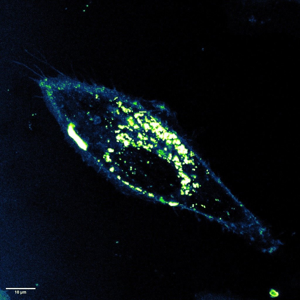
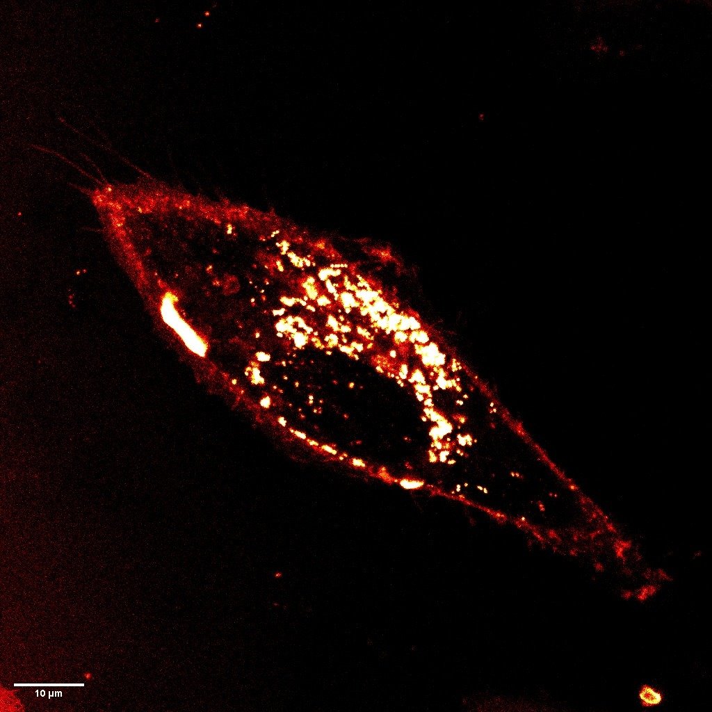
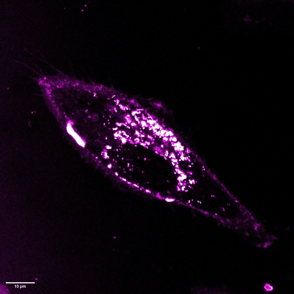
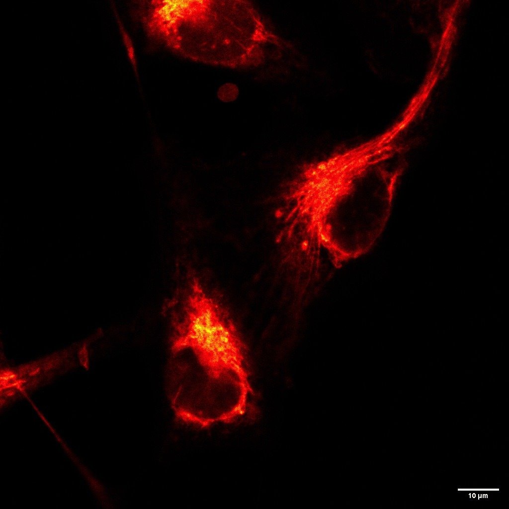
Imaging of H28-living cells stained with far-red LBL-Dye CellTag 717 (5 µg/µl) in the culture medium. Images acquired by using SP5 confocal microscope (Leica Microsystems), X63 objective oil immersion, WLL laser excitation 690 nm; Em 700-730 nm. The plasma membrane and vesicles are visible on living cells and the movement of cells can be followed during few hours.

The efficacy of fluorescence-guided surgery in facilitating the real-time delineation of tumours depends on the optical contrast of tumour tissue over healthy tissue. Here we show that CJ215—a commercially available, renally cleared carbocyanine dye sensitive to apoptosis, and with an absorption and emission spectra suitable for near-infrared fluorescence imaging (wavelengths of 650–900 nm) and shortwave infrared (SWIR) fluorescence imaging (900–1,700 nm)—can facilitate fluorescence-guided tumour screening, tumour resection and the assessment of wound healing...
The efficacy of fluorescence-guided surgery in facilitating the real-time delineation of tumours depends on the optical contrast of tumour tissue over healthy tissue. Here we show that CJ215—a commercially available, renally cleared carbocyanine dye sensitive to apoptosis, and with an absorption and emission spectra suitable for near-infrared fluorescence imaging (wavelengths of 650–900 nm) and shortwave infrared (SWIR) fluorescence imaging (900–1,700 nm)—can facilitate fluorescence-guided tumour screening, tumour resection and the assessment of wound healing...
The efficacy of fluorescence-guided surgery in facilitating the real-time delineation of tumours depends on the optical contrast of tumour tissue over healthy tissue. Here we show that CJ215—a commercially available, renally cleared carbocyanine dye sensitive to apoptosis, and with an absorption and emission spectra suitable for near-infrared fluorescence imaging (wavelengths of 650–900 nm) and shortwave infrared (SWIR) fluorescence imaging (900–1,700 nm)—can facilitate fluorescence-guided tumour screening, tumour resection and the assessment of wound healing...
Proimaging